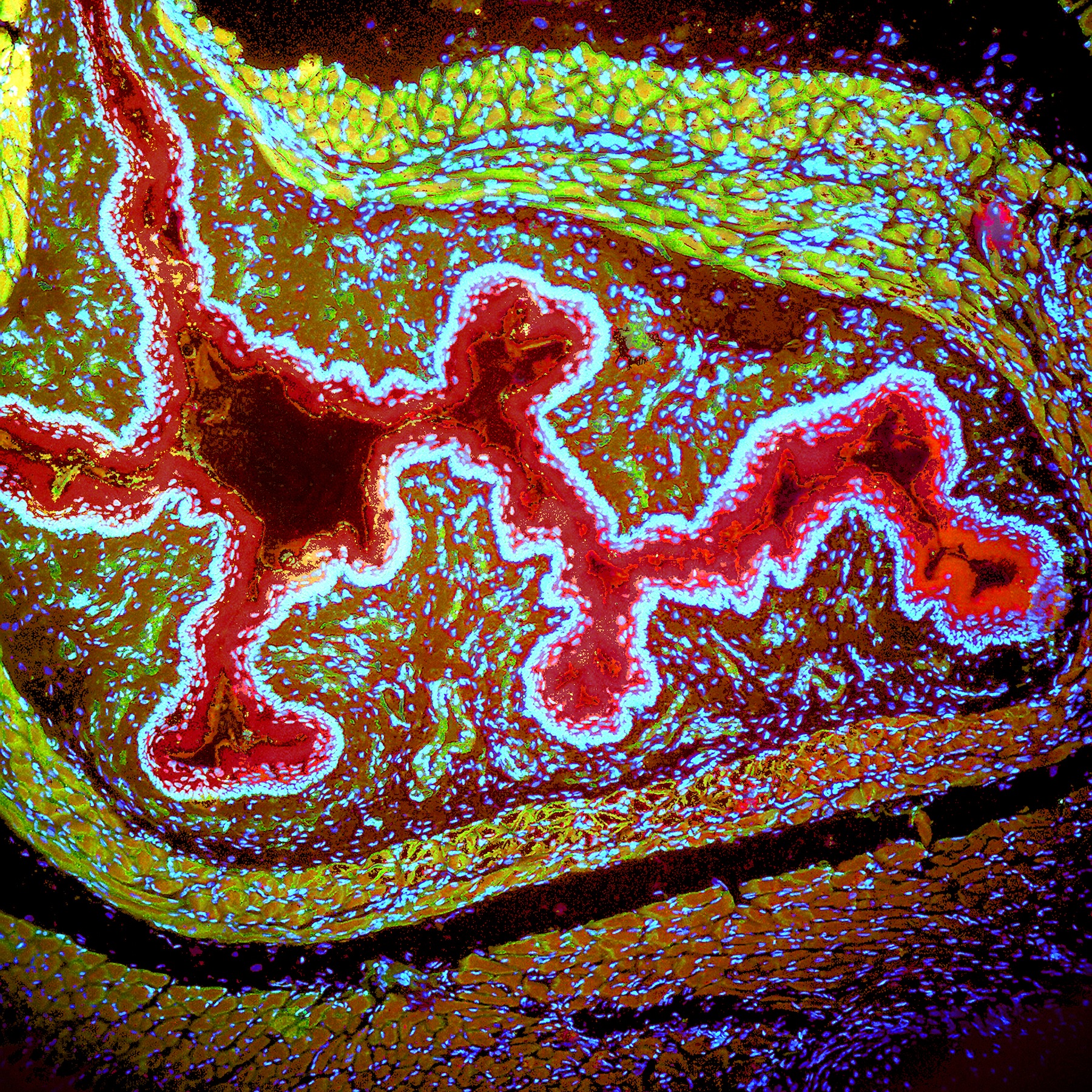
A cross section of rat trachea stained with three different fluorescent dyes, each of which has an affinity for different tissues/molecules and emits a different wavelength of light; Hoechst (blue), Texas red, and FITC (green). The blue spots are the DNA in the nucleus of each cell in the tissue.
The College of the Canyons Art Gallery will fuse the aesthetics of fine art and photography with the often invisible beauty of the natural world to create a first-of-its-kind presentation with the exhibition “Life As Art: Images From an Unseen World.”
As the culmination of a multi-year collaboration between COC biology professors Kelly Burke and Kelly Cude, this exhibition includes more than 60 micrographs (photographs taken with a microscope) produced while working in the college’s biology laboratories.
“I am very proud of this marvelous collaboration between the Art Gallery, the biology department andthese two terrific instructors,” said Larry Hurst, director of the COC Art Gallery. “I think that the community will agree that this show represents a wonderful partnership between the visual arts and the life sciences.”
Designed to introduce viewers to depictions of the natural world that are rarely, if ever, seen, this exhibition will amaze audiences with its array of abstract, sophisticated and microscopically enhanced images of real world objects.
“To me this show is an opportunity for people to change their perspective of the world around them by experiencing the intrinsic beauty and complexity of the microscopic world,” said Cude. “When people realize that an entire microscopic universe can exist in a single drop of pond water, or that humans are made up of trillions and trillions of independent living cells, they cannot help but be amazed and have a deeper appreciation for life.”
Micrographs on display will include artful depictions of plants, animals and microbes, as well as a variety of human cells and tissues. Many of the images come from the college’s biological slide and specimen collection. Others were specifically cultured and prepared, or collected from natural environments.
“My goal for this project was to create images of the microscopic world that I love so much, and attempt to share that beauty with others,” said Burke. “To my surprise, I’ve gained a new appreciation for abstract representation.”
The exhibition “Life as Art: Images From an Unseen World” will be on display at the COC Art Gallery from Monday, Aug. 26 through Thursday, Sept. 26, 2013.
An artist reception will take place in the Art Gallery from 2 to 4 p.m. Saturday, Sept. 21.
The College of the Canyons Art Gallery is open 11 a.m. to 3 p.m. Monday through Thursday. Those unable to visit the gallery during normal hours are welcome to contact the gallery to schedule a viewing appointment.
All gallery exhibitions and related events are free and open to the public.
For more information about the College of the Canyons Art Gallery or the exhibition “Life as Art: Images From an Unseen World visit www.canyons.edu/artgallery.

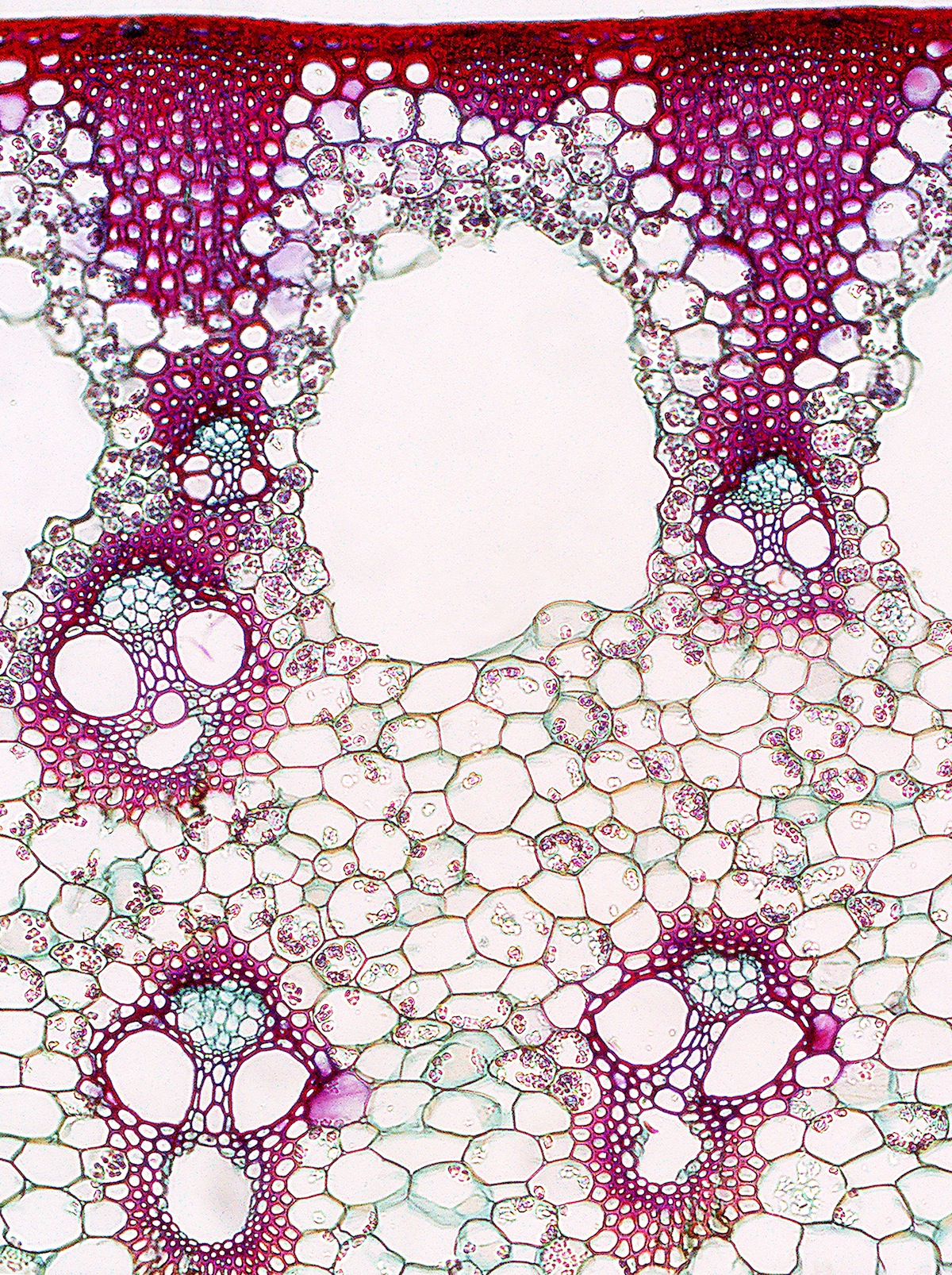
Vascular bundles in stem section imaged using bright field microscopy. Magnification is 100X. Average diameter of vascular bundle is 0.17mm. Seeing this “face” pop up under the microscope did surprise me! It is an image of a non-woody plant stem showing the two main types of vascular tissue: (1) xylem which carries nutrients and water up from the roots to the rest of the plant and (2) phloem which carries sugars and nutrients generated during photosynthesis down to all parts of the plant. The “tube-like” networks of xylem and phloem are bundled together into arrangements known as vascular bundles, which resemble “miniature faces.”

Cultured human neurons imaged using fluorescent microscopy. Magnification is 1000X. Neurons are 0.02 to 0.05 mm in length. These neurons were grown attached to petri dishes inside incubators that mimic human body temperature and humidity. Cells were then stained with a fluorescent green antibody that binds onto the cell’s cytoskeleton (“skeleton fibers of the cell”). This outlines the nerve cell body and nerve projections (axons and dendrites) in bright green, while leaving the nucleus, which is devoid of cytoskeleton, as a black area inside each cell body.
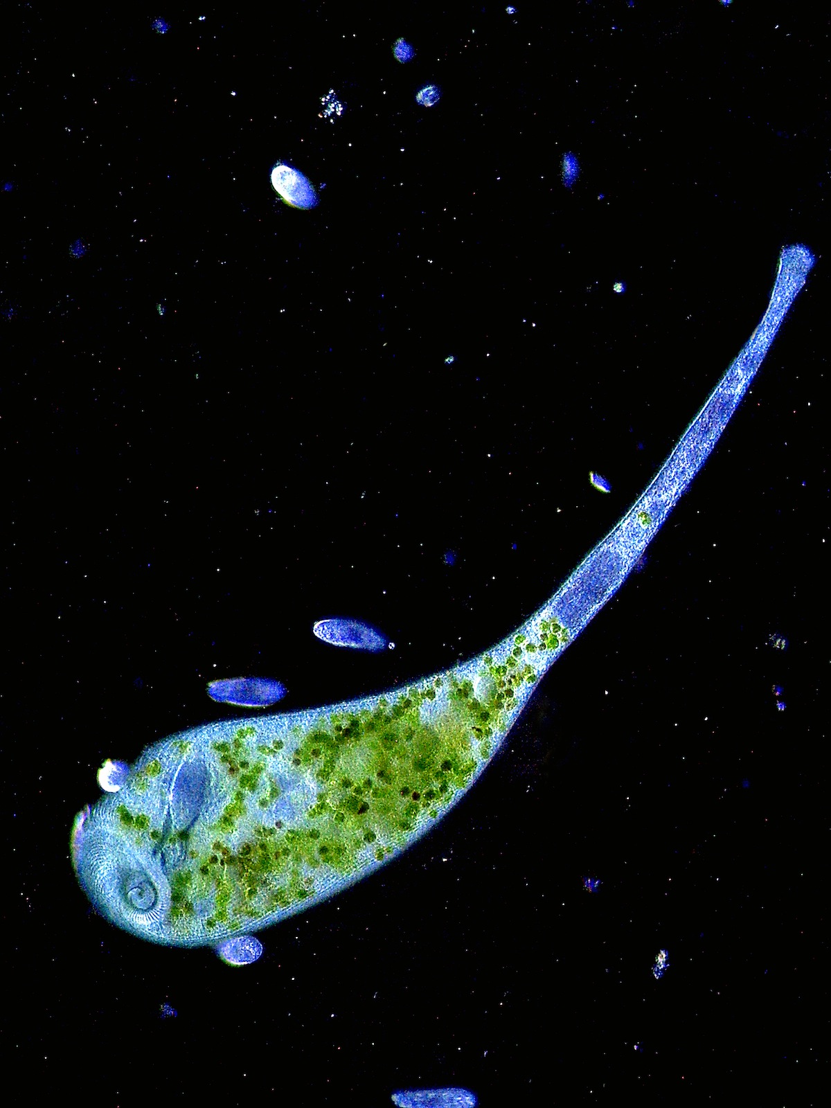
This completely extended Stentor (protozoa) cell shows the complexity that can be found in a microscopic single celled organism. The trumpet opening is lined with cilia which facilitate feeding as food items spiral into the gullet; algae which will live inside the Stentor producing carbohydrates via photosynthesis which the Stentor can then utilize.
Vascular bundles in stem section imaged using bright field microscopy. Magnification is 100X. Average diameter of vascular bundle is 0.17mm. Seeing this “face” pop up under the microscope did surprise me! It is an image of a non-woody plant stem showing the two main types of vascular tissue: (1) xylem which carries nutrients and water up from the roots to the rest of the plant and (2) phloem which carries sugars and nutrients generated during photosynthesis down to all parts of the plant. The “tube-like” networks of xylem and phloem are bundled together into arrangements known as vascular bundles, which resemble “miniature faces.”
Aug. 26: 'Images From an Unseen World' Opens at COC Art Gallery